November 22, 2013
MyFootShop.com
Venous Stasis Dermatitis and Venous Ulcerations
- Summary
- Symptoms
- Read More
Summary
Stasis dermatitis (also called venous stasis dermatitis) is an inflammatory reaction of the skin of the lower leg caused by static (slow or delayed) venous flow of the leg. Stasis dermatitis is common in patients 50 years and older. Left untreated, venous stasis can progress to a venous ulceration and lower leg infection. The most common location for a venous ulceration of the lower extremity is over the medial aspect of the ankle. Venous ulcerations vary in size and depth. Stasis dermatitis and venous ulcerations are more common in women than men.
Symptoms
- Redness, dryness, and peeling of the distal 1/3 to 1/2 of the lower leg
- Firm swelling of the distal leg
- Pitting edema of the foot and leg
- Ulceration of the medial ankle
The symptoms of stasis dermatitis vary with the general health of the patient, duration of the condition, and status of treatment. Early stasis dermatitis shows a red blush surrounding the lower 1/3 of the leg. Stasis dermatitis is usually found in both legs with symmetrical presentation. Pigmentation changes (darkening) of the leg usually follows a prolonged period of swelling (edema) of the lower leg.
Description
 Circulation describes the flow of blood from the heart to the toes and back to the heart, making a circle or loop. The circulation circle has two sides that include arterial and venous circulation. Arterial blood is oxygenated and moves away from the heart to the extremities while venous blood is poorly oxygenated and returns to the heart. Venous stasis describes the slowing of venous blood flow in the lower leg as blood returns from the foot to the heart. This slowing of venous blood flow in the legs is commonly caused by incompetent valves within the veins of the leg. The inability of blood to flow effectively up the leg results in swelling of the lower leg (venous hypertension) and a visible change in the
  skin of the lower leg called venous stasis dermatitis. As the duration of edema continues, stasis dermatitis will worsen with eczematization and lichenification of the leg. Eczematization is the thickening of the skin. Lichenification describes crusting and layering of the skin that occurs when the normal exfoliative properties of the skin are disrupted by chronic swelling. In severe cases of stasis dermatitis, oozing, scaling, and deep pigmentation changes in the skin may become apparent. Chronic venous stasis dermatitis results in the skin and deeper tissues of the lower leg becoming indurated (firm.)
If left untreated, stasis dermatitis will progress to where the skin ulcerates due to venous hypertension caused by venous valvular incompetency. In this stage, the leg will open and weep fluid (serum and extracellular fluid). Infection may also become a secondary issue with both venous stasis dermatitis and venous ulcerations.

The function of the venous system of the leg is to return blood from the foot back to the heart. The lower extremity venous system consists of three types of veins; superficial, perforator and deep veins. Veins are very similar to arteries, with the exception that veins have less muscle and carry a lower intra-luminal pressure. Superficial and deep veins contain valves that open with blood flow to the heart and close with a reversal of flow. The purpose of the valves is to ensure unidirectional flow of blood to the heart. The veins of the leg are unique in that they also use muscular contraction of the lower leg to drive blood to the heart. This pump is called the calf pump. While walking, compression of the vein by the muscles of the lower leg helps to facilitate blood flow to the heart.
The two primary superficial veins of the leg are the great saphenous and the small saphenous veins. While standing, the pressure in each of these veins is equal to the pressure that would be found in a column of blood extending from the foot to the heart. When the calf pump is activated, blood is pumped proximally through the perforator veins into the deep veins. The deep veins of the leg include the posterior tibial vein, anterior tibial vein, popliteal vein, and femoral vein.
Causes and contributing factors
Factors that can contribute to the early onset of stasis dermatitis include obesity, inactivity, venous injury, dependency (lower than the heart) of the leg and infection of the leg. The primary contributing cause of stasis dermatitis is valvular incompetency of the veins of the leg resulting in chronic edema (swelling.)
Differential diagnosis
The differential diagnosis for venous stasis includes:
Cellulitis
Peripheral arterial disease
Psoriasis
Varicose veins
Treatment
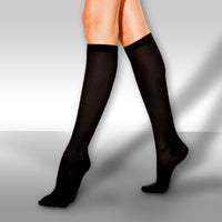
Central to the treatment of venous stasis dermatitis and venous stasis ulcers is control of lower extremity edema due to venous hypertension. Edema can be controlled by elevating the legs above the level of the heart, use of diuretics and the use of compression hose. It's important to realize that when using compression hose, the hose needs to be put on first thing in the morning in advance of any swelling. If swelling is allowed to occur before applying support hose, that swelling will likely be there for the rest of the day. Additional tools used to manage peripheral edema include edema pumps, compression dressings, and a Circ-Aid.
Eczematous changes (peeling and flaking) and lichenification (hardening of the epidermis) can be treated with skin softening agents. Inflammatory changes of the skin are common and can be treated with topical or oral steroids. Discoloration of the skin is difficult to treat. Discoloration or darkening of the skin is often due to the deposition of hemosiderin (the iron component of red blood cells). Once hemosiderin is deposited in the skin, it is much like a tattoo that stains the skin from within and is permanent.
Venous ulceration of the skin is common, particularly at the medial (inside) ankle. The area superficial to the origin of the great saphenous vein is the most common site of ulceration. Ulcerations should be cultured and treated for infection if necessary. Compression boots are the gold standard for treating venous stasis ulcerations. Compression boots can be made from a number of materials but the primary role of the compression boot is to manage peripheral edema. Compression boots are changed on a weekly basis. Treatment can take from one to many weeks to see complete closure of the venous ulcer. Negative pressure treatment of the wound along with skin grafting may be necessary in severe, non-healing ulcers.
Surgical treatment of venous stasis and recurrent venous legs wounds is often performed to prevent recurrence. Treatment consists of radiofrequency ablation of the superficial veins and excision of perforating veins of the lower leg. Surgical correction of recurrent venous wounds does not preclude the need to wear compression hose.
Care of venous stasis ulcerations can be complex and is best managed by a team of specialists at a wound care center. Advanced treatment modalities are difficult to manage in an office setting, therefore, wound care centers often become an important part of care with venous stasis ulcerations.
When to contact your doctor
Management of venous stasis and venous ulcerations should not be attempted at home. Consult your podiatrist or local wound care center for additional treatment recommendations.
ICDM-10 Codes - Venous stasis dermatitis and venous ulcerations
(I87.8) Venous hypertension
(I87.8) Venous stasis
(I83.001) Varicose veins of unspecified lower extremity with ulcer of thigh
(I83.002) Varicose veins of unspecified lower extremity with ulcer of calf
(I83.003) Varicose veins of unspecified lower extremity with ulcer of ankle
(I83.004) Varicose veins of unspecified lower extremity with ulcer of heel and midfoot
(I83.005) Varicose veins of unspecified lower extremity with ulcer other part of foot
(I83.008) Varicose veins of unspecified lower extremity with ulcer other part of lower leg
(I83.009) Varicose veins of unspecified lower extremity with ulcer of unspecified site
(I87.311) Chronic venous hypertension (idiopathic) with ulcer of right lower extremity
(I87.312) Chronic venous hypertension (idiopathic) with ulcer of left lower extremity
(I87.313) Chronic venous hypertension (idiopathic) with ulcer of bilateral lower extremity
(I87.319) Chronic venous hypertension (idiopathic) with ulcer of unspecified lower extremity
(I87.331) Chronic venous hypertension (idiopathic) with ulcer and inflammation of right lower extremity
(I87.332) Chronic venous hypertension (idiopathic) with ulcer and inflammation of left lower extremity
(I87.333) Chronic venous hypertension (idiopathic) with ulcer and inflammation of bilateral lower extremity
(I87.339) Chronic venous hypertension (idiopathic) with ulcer and inflammation of unspecified lower extremity
(L97.901) Non-pressure chronic ulcer of unspecified part of unspecified lower leg limited to breakdown of skin
(L97.902) Non-pressure chronic ulcer of unspecified part of unspecified lower leg with fat layer exposed
(L97.903) Non-pressure chronic ulcer of unspecified part of unspecified lower leg with necrosis of muscle
(L97.904) Non-pressure chronic ulcer of unspecified part of unspecified lower leg with necrosis of bone
(L97.905) Non-pressure chronic ulcer of unspecified part of unspecified lower leg with muscle involvement without evidence of necrosis
(L97.906) Non-pressure chronic ulcer of unspecified part of unspecified lower leg with bone involvement without evidence of necrosis
(L97.908) Non-pressure chronic ulcer of unspecified part of unspecified lower leg with other specified severity
(L97.909) Non-pressure chronic ulcer of unspecified part of unspecified lower leg with unspecified severity
(L97.911) Non-pressure chronic ulcer of unspecified part of right lower leg limited to breakdown of skin
(L97.912) Non-pressure chronic ulcer of unspecified part of right lower leg with fat layer exposed
(L97.913) Non-pressure chronic ulcer of unspecified part of right lower leg with necrosis of muscle
(L97.914) Non-pressure chronic ulcer of unspecified part of right lower leg with necrosis of bone
(L97.915) Non-pressure chronic ulcer of unspecified part of right lower leg with muscle involvement without evidence of necrosis
(L97.916) Non-pressure chronic ulcer of unspecified part of right lower leg with bone involvement without evidence of necrosis
(L97.918) Non-pressure chronic ulcer of unspecified part of right lower leg with other specified severity
(L97.919) Non-pressure chronic ulcer of unspecified part of right lower leg with unspecified severity
(L97.921) Non-pressure chronic ulcer of unspecified part of left lower leg limited to breakdown of skin
(L97.922) Non-pressure chronic ulcer of unspecified part of left lower leg with fat layer exposed
(L97.923) Non-pressure chronic ulcer of unspecified part of left lower leg with necrosis of muscle
(L97.924) Non-pressure chronic ulcer of unspecified part of left lower leg with necrosis of bone
(L97.925) Non-pressure chronic ulcer of unspecified part of left lower leg with muscle involvement without evidence of necrosis
(L97.926) Non-pressure chronic ulcer of unspecified part of left lower leg with bone involvement without evidence of necrosis
(L97.928) Non-pressure chronic ulcer of unspecified part of left lower leg with other specified severity
(L97.929) Non-pressure chronic ulcer of unspecified part of left lower leg with unspecified severity
The International Classification of Diseases, Tenth Revision (ICD-10) was developed and is maintained by the World Health Organization (WHO). The U.S. National Center for Health Statistics (NCHS) is the WHO Collaborating Center for the Family of International Classifications in North America, and is the U.S. governmental agency responsible for overseeing ICD use in the U.S.
CPT Codes - Venous stasis and venous ulcerations
(11042) Debridement, subcutaneous tissue (includes epidermis and dermis, if performed); first 20 sq cm or less
(11043) Debridement, muscle and/or fascia (includes epidermis, dermis, and subcutaneous tissue, if performed); first 20 sq cm or less
(11045) Debridement, subcutaneous tissue (includes epidermis and dermis, if performed); each additional 20 sq cm, or part thereof (List separately in addition to code for primary procedure)
(29580) Strapping; Unna boot
(29581) Application of multi-layer compression system; leg (below knee), including ankle and foot
(97597) Debridement (eg, high-pressure waterjet with/without suction, sharp selective debridement with scissors, scalpel, and forceps), open wound, (eg, fibrin, devitalized epidermis and/or dermis, exudate, debris, biofilm), including topical application(s), wound assessment, use of a whirlpool, when performed and instruction(s) for ongoing care, per session, total wound(s) surface area; first 20 sq cm or less
(97598) Debridement (eg, high-pressure waterjet with/without suction, sharp selective debridement with scissors, scalpel, and forceps), open wound, (eg, fibrin, devitalized epidermis and/or dermis, exudate, debris, biofilm), including topical application(s), wound assessment, use of a whirlpool, when performed and instruction(s) for ongoing care, per session, total wound(s) surface area; each additional 20 sq cm, or part thereof (List separately in addition to code for primary procedure)
(97602) Removal of devitalized tissue from wound(s), non-selective debridement, without anesthesia (eg, wet-to-moist dressings, enzymatic, abrasion), including topical application(s), wound assessment, and instruction(s) for ongoing care, per session
(15430) Acellular xenograft implant; first 100 sq cm or less, or 1% of body area of infants and children
(15431) Acellular xenograft implant; each additional 100 sq cm, or each additional 1% of body area of infants and children, or part thereof (List separately in addition to code for primary procedure)
(Q4102) Oasis wound matrix, per square centimeter
(Q4124) Oasis ultra tri-layer wound matrix, per square centimeter
(C9365) Oasis ultra tri-layer matrix, per square centimeter
Current Procedural Terminology (CPT®) is a product of the American Medical Association (AMA).
References
1. London NJ, Donnelly R. ABC of arterial and venous disease. Ulcerated lower limb. BMJ. 2000;320:1589–91.
2. Nelzén O, Bergqvist D, Lindhagen A. Venous and non-venous leg ulcers: Clinical history and appearance in a population study. Br J Surg. 1994;81:182–7.
Author(s) and date
![]() This article was written by Myfootshop.com medical advisor Jeffrey A. Oster, DPM.
This article was written by Myfootshop.com medical advisor Jeffrey A. Oster, DPM.
Cite this article as: Oster, Jeffrey. Venous Stasis Dermatitis and Venous Ulcerations. /blogs/articles/venous-stasis-dermatitis-and-venous-ulcerations
Most recent article update: January 15, 2021.
 Venous Stasis Dermatitis and Venous Stasis Ulcers by Myfootshop.com is licensed under a Creative Commons Attribution-NonCommercial 3.0 Unported License.
Internal use only: ZoneD2
Filed in:
category-leg,
category-other,
category-skin-nail,
circulation problems,
cmsid-154,
image-1506,
skin problems,
venous stasis dermatitis,
venous ulceration,
wounds of the leg,
ZoneD2