Peroneal Tendon Rupture
- Summary
- Symptoms
- Read More
Summary
Peroneal tendon ruptures are often the result of an inversion ankle sprain. During an ankle sprain, the peroneal tendons pull up against the outside of the ankle to restrain the rolling motion of the ankle. The force applied to the peroneal tendons can be enough to create a tear (rupture) of the tendon. Most tears of the peroneal tendons are partial ruptures of the peroneus brevis tendon called longitudinal tears, tearing along the length of the tendon. Peroneal tendon tears are found equally between men and women. The onset of tears are during adult working years, ages 25 to 60.
Symptoms
- Chronic lateral ankle pain following a simple inversion sprain
- Chronic swelling of the posterior lateral ankle
- Crepitus (palpable crackle) posterior aspect of the lateral ankle with range of motion
- Pain with ambulation, particularly during the contact and push off phase of gait
- Increased posterior lateral ankle pain with toe raises
Description
The peroneal muscles and tendons have two functions. Their primary function is to stabilize the foot as the body passes over the foot. The peroneal muscles and tendons will help to stabilize the foot on uneven, rough surfaces. As the foot rolls from side to side, the peroneal muscles and tendons help to inhibit a lateral ankle sprain by preventing the foot from rolling to the outside (lateral side) of the foot. The secondary function of the peroneal muscles and tendons is to assist the calf with plantarflexion.
During an ankle sprain, as the ankle begins to roll, the peroneal muscles fire to stabilize the ankle and prevent the lateral ankle sprain from occurring. Both tendons are pulling up against the downward force of the lateral ankle. The fibula (lateral ankle bone) becomes a wedge, carrying body weight down toward the ground. As the fibula drives downward, the peroneal tendons pull up against the ankle and are compressed. The peroneus brevis tendon is adjacent to the fibula while the peroneus longus tendon runs external to the brevis tendon. A longitudinal tear occurs when the peroneus longus tendon pulls so hard that it transects (slices) the brevis tendon into two parts along its length. This means that the injury is actually caused by one peroneal tendon (the longus) transecting the other peroneal tendon (the brevis.)
The peroneus longus tendon is not immune to injury. Partial and complete ruptures of the peroneus longus tendon do occur but are far less common than injuries seen in the peroneus brevis tendon. The weakest portion of the peroneus longus tendon is the point where it changes direction and rounds the plantar surface of the cuboid. When ruptures of the peroneus longus do occur, they tend to be found just distal to the plantar cuboid and are also longitudinal. Complete transverse ruptures of the peroneus longus tendon are rare.
Another uncommon injury of the peroneus longus tendon is the rupture of the tendon at the site of an os peroneum. The os peroneum is a small accessory bone found within the peroneus longus tendon at the lateral wall of the cuboid. The occurrence of an os peroneum in the general population is reported in the literature to be 5-26%. When present, a healthy, functioning os peroneum will help facilitate the transfer of load carried by the peroneus longus as it rounds the cuboid. Bipartite (two part) os peroneum are common. Bipartite os peroneum and fractured os peroneum can be difficult to differentiate. When viewed on x-ray, a bipartite os peroneum will typically have smooth edges while a fractured os peroneum will display ragged edges. MRI is helpful in differentiating between a bipartite or fractured os peroneum.
The peroneal tendons originate high in the lateral aspect of the leg attaching to both the tibia and fibula. They descend the lateral compartment of the leg rounding the posterior aspect of the lateral malleolus (outside ankle bone or fibula) to continue down to the lateral side of the foot. The peroneus brevis inserts into the base of the fifth metatarsal. The peroneus longus curves under the midfoot to the bottom of the arch. Both the brevis and longus are innervated by the superficial peroneal nerve.
Causes and contributing factors
Peroneal tendon tears are directly related to trauma to the foot or ankle. Any biomechanical characteristic of the foot that may contribute to an ankle sprain may contribute to a peroneal tendon tear. Those biomechanical characteristics may include rearfoot varus, forefoot valgus or metatarsus adductus.
Differential diagnosis
The differential diagnosis for a peroneal tendon tear includes:
Achilles tendinitis
Achilles tendon rupture
Ankle sprain
Arthritis
Broken ankle
Calcaneal stress fracture
Peroneal tendinitis
Retrocalcaneal bursitis
Sinus tarsitis
Treatment
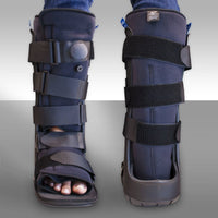
 Initial care of peroneal tendon ruptures includes much of the same care recommended for ankle sprains: rest, ice, elevation, compression, and anti-inflammatory medications. A 4-6 week period of conservative care is warranted post-ankle sprain before obtaining further testing such as an MRI. Use of a walking cast or ankle brace may help to splint the peroneal tendons during conservative care. Most peroneal brevis tendon ruptures do not heal with mere conservative care and will require surgical repair.
Following a lateral ankle sprain, if the lateral ankle is still painful at 6 weeks post-injury, an MRI may help to determine whether the peroneal tendons have sustained an injury. Alternatively, diagnostic ultrasound may be used to evaluate partial ruptures of the peroneal tendons. MRI is not always 100% accurate when evaluating peroneal tendon pathology. Many cases of peroneal tears are too small to find with an MRI or ultrasound and can only be found with direct visualization during surgery. Occasionally, an accessory tendon known as the peroneus tertius is present within the peroneal tendon sheath and is misdiagnosed on MRI as a tendon tear.
The following images show the steps used to perform a repair of a severe longitudinal tear of the peroneus brevis tendon. Image 1 shows pre-operative planning outlining the leg and fibula to the left along with the 5th metatarsal and toes to the right. Image 2 shows dissection through the subcutaneous space and entry into the combined sheath of the peroneal tendons. Image 3 shows the initial appearance of the damaged peroneus brevis tendon. Image 4 shows the dissection of the injury in greater detail. The peroneus brevis tendon shows myxoid degeneration (scaring) and multiple tears. Image 5 shows an intact peroneus longus tendon with mildly reactive synovium lining the inside wall of the peroneal tendon sheath. This reaction is due to chronic inflammation within the tendon sheath. Image 6 shows the repaired peroneus brevis tendon. Also very clear in this image is the peroneal retinaculum. Image 7 shows final skin closure.


Surgical repair of a longitudinal peroneus brevis tear can be performed on an outpatient basis using sedation and local anesthesia or general anesthesia. The procedure takes approximately 45 minutes to complete. Following repair, most doctors will limit ambulation to partial weight bearing for a period of days to weeks. No casting is necessary as early non-weight bearing range of motion is desired. Return to normal activates depends upon the severity of the tear and success of the surgery. Most patients are back to 75% of normal activities by 4 weeks post-surgery.
In severe cases of peroneus brevis or peroneus longus tears, including complete ruptures, treatment options do vary. Tenodesis (fixation of the tendon) of the damaged tendon may be completed by permanently attaching the tendon to the cuboid, calcaneus or adjacent tendon. For instance, in cases of severe peroneus brevis ruptures, the peroneus brevis tendon may be permanently attached (tenodesed) to the peroneus longus tendon. Other options include the use of supplemental graft material such as a split thickness graft from the Achilles tendon of synthetic grafting material.
In cases of a symptomatic os peroneum or fractured os peroneum, the majority of cases can be resolved with simple excision of the os peroneum. Excision of the os peroneum can be performed with a general anesthetic or a local anesthetic with sedation. Recovery varies and depends upon the integrity of the peroneus longus tendon following the surgery. The peroneus tendon will be weakened by excision of the os peroneum but will regain full strength over several months. Limitations on ambulation post-surgery depend upon the surgeon's impression of the status of the tendon post-op. Limitations may include non-weight bearing or partial weight bearing for a period of 6-8 weeks post-op.
When to contact your doctor
Any lateral ankle injury that fails to respond in 6-8 weeks to conservative care should be evaluated by your podiatrist or orthopedist to rule out a peroneal tendon tear.
References
References are pending.
Author(s) and date
![]() This article was written by Myfootshop.com medical advisor Jeffrey A. Oster, DPM.
This article was written by Myfootshop.com medical advisor Jeffrey A. Oster, DPM.
Competing Interests - None
Cite this article as: Oster, Jeffrey. Peroneal Tendon Rupture. /blogs/articles/peroneal-tendon-rupture
Most recent article update: January 14, 2021.
 Peroneal Tendon Rupture by Myfootshop.com is licensed under a Creative Commons Attribution-NonCommercial 3.0 Unported License.
Internal reference only: ZoneP4, ZoneD3, ZoneL3