Posterior Tibial Tendon Dysfunction
- Summary
- Symptoms
- Read More
- Video
Summary
Posterior tibial tendon dysfunction (PTTD) is the leading cause of acquired flatfoot in adults. The stages of PTTD include inflammation of the tendon, partial tear or complete tendon tear. The onset of PTTD is usually insidious and the condition is progressive over time. An abrupt onset is possible but is typically linked to some form of trauma, whether it be simple (stepping down off a curb or ladder) or severe (falling from a height or automobile accident.) PTTD is seldom seen in children and increases in frequency with age. The onset of PTTD is unilateral but it is not uncommon to see bilaterally. Posterior tibial tendon dysfunction is found equally in men and women.
Symptoms
- Loss of medial arch height
- Painful swelling of the medial arch
- Pain increased with weight bearing and toe raise
- Often lateral ankle pain found in addition to medial arch pain
Description
 There have been many proposed explanations for posterior tibial tendon dysfunction (PTTD) over the years since this condition was first described by Kulkowski in 1936. The most contemporary explanation refers to an area of hypovascularity (limited blood flow) in the tendon just below the ankle. The tendon derives most of its nutritional support from synovial fluid produced by the inner lining of the tendon. Extremely small blood vessels also permeate the tendon sheath to reach the tendon. This anatomical limitation makes all tendons notoriously slow to heal. In the case of the posterior tibial tendon, this problem is exacerbated by a distinct area of poor blood flow (hypovascularity). This area of hypovascularity is located in the posterior tibial tendon just below or distal to the inside ankle bone (medial malleolus.)
PTTD is a progressive condition, meaning to say that if left untreated, PTTD will worsen over time. The onset of PTTD begins with focal tendinitis. If left untreated, tendinitis will progress to partial and then complete tears of the posterior tibial tendon. As PTTD becomes more severe, the ability of the posterior tibial tendon become less able to support the arch. Hence the collapse of the arch associated with stage III PTTD. Several classifications have been developed to describe posterior tibial tendon dysfunction. The classification as described by Johnson and Strom is most commonly used today.[1,2]
Stage I Posterior tibial tendonitis without tendon tear:
Tendon status - Attenuated (lengthened) with tendinitis but no rupture.
Clinical findings - Palpable pain in the medial arch. Foot is supple, flexible. Too many toes sign may be positive or negative.
X-ray/MRI - Mild to moderate tenosynovitis on MRI, no X-ray changes found.
Stage II Posterior tibial tendonitis with partial tendon tear:
Tendon status - Attenuated with possible partial or complete rupture.
Clinical findings - Pain in arch. Unable to raise on toes. Too many toes sign positive.
X-ray/MRI - MRI notes tear in tendon. X-ray noting abduction of forefoot, collapse of talo-navicular joint.
Stage III Posterior tibial tendonitis with partial to complete tendon tear:
Tendon status - Severe degeneration of the tendon with likely rupture.
Clinical findings - Rigid flatfoot with inability to raise up on toes. Too many toes sign positive.
X-ray/MRI - MRI shows tear in tendon. X-ray noting abduction of forefoot, collapse of talo-navicular joint.
The posterior tibial tendon (tibialis posterior) is the extension of the posterior tibial muscle that lies deep to the calf. The origin of the posterior tibial muscle is the posterior aspect of both the tibia and fibula and the interosseous membrane. The insertion of the posterior tibial muscle is the medial navicular, where the tendon divides into nine different insertion sites on the bottom of the foot. The function of the posterior tibial tendon is to plantarflex the foot at the toe off phase of the gait cycle and to stabilize the medial arch and subtalar joint as the body passes over the foot.
Causes and contributing factors
The prevalence of adult-onset PTTD increases with age and is found in 3.3% of postmenopausal women over the age of 40.[3] The literature doesn't clearly differentiate a predilection for PTTD in women or men. This article focuses on progressive, adult-onset PTTD in the non-athletic population.
An early onset form of PTTD can also be found in younger athletes and is often referred to as posterior shin splints. Posterior shin splints can be defined as stage 1 PTTD. [4,5]
The tendon is most susceptible to fatigue and failure at an area where the tendon changes direction. As the posterior tibial tendon descends the leg and descends to the inner ankle, the tendon follows a well-defined groove in the back of the tibia (bone of the inside of the ankle). The tendon then takes a dramatic turn towards the arch of the foot. If the tendon is put into a situation where significant load is applied to the foot, the tendon responds by pulling up as the load of the body (in addition to gravity) pushes down. At the location where the tendon changes course, the tibia acts as a wedge and may apply enough force to actually damage or rupture the tendon.
Equinus is also a contributing factor in cases of PTTD. Equinus is the term used to describe the ability or lack of ability to dorsiflex the foot at the ankle (move the toes towards the shin). Equinus is usually due to tightness in the calf muscle, also known as the gastroc-soleal complex (a combination of the gastrocnemius and soleus muscles). Equinus may also be due to a bony block in the front of the ankle. The presence of equinus forces the posterior tibial tendon to accept additional load during gait.
Additional contributing factors to the onset of posterior tibial tendon dysfunction may include obesity, hypertension, diabetes, peripheral neuropathy, smoking or arthritis. Although PTTD is often associated with peri or post-menopausal women, no evidence exists to suggest that PTTD is hormonal-based.[6]
Differential diagnosis
The differential diagnosis for PTTD includes:
Arthritis
Anterior tibialis tendinitis
Calcaneal stress fracture
Flexor hallucis longus tendonitis
Posterior tibial tendon rupture
Shepard's fracture
Sinus tarsi syndrome
Subtalar joint arthritis
Symptomatic os tibiale externum
Tarsal tunnel syndrome
Tibial stress fracture
 The os tibiale externum, or what is frequently called and accessory navicular, is a small bone that resides within the body of the PT tendon. The os tibiale externum functions to facilitate motion around the navicular. The os tibiale externum functions much in the same way that the knee cap (patella) works to guide the quadraceps tendon around the knee as it bends. The os tibiale externum can undergo degenerative wear called chondromalacia. The os tibiale externum also can fracture. Therefore, the os tibiale externum must also be considered when diagnosing PT tendon pain and PTTD.
Treatment
The diagnosis of PTTD is made by a combination of patient history, clinical exam, x-ray, ultrasound, and MRI. The clinical exam includes a non-weight bearing and weight bearing exam of the foot. X-rays are used to identify any secondary contributing factors such as boney blocks of the foot or arthritis. Ultrasound or MRI are very useful to assess the integrity of the posterior tibial tendon.
A common test to evaluate PTTD is the "too many toes sign." The "too many toes sign" is a test used to measure abduction (deviation away from the midline of the body) of the forefoot. With damage to the posterior tibial tendon, the forefoot will abduct or move out in relationship to the rest of the foot. In cases of PTTD, when the foot is viewed from behind, the toes appear as "too many" on the outside of the foot due to abduction of the forefoot.
Treatment of PTTD is dependent upon the clinical stage and the health status of the patient.[2] It is important to recognize that PTTD is a mechanical problem that requires a mechanical solution. This means that treating PTTD with medication alone is fraught with failure. Prompt introduction of some form of mechanical support is imperative.[7,8]
Surgical procedures which focus on primary repair of the posterior tibial tendon by itself have proven to be very unsuccessful. This is due to the fact that tendon heals slowly following injury and cannot be relied upon as a sole solution for PTTD cases. Surgical success is usually achieved by stabilization of the rearfoot (subtalar joint) which significantly reduces the work performed by the posterior tibial tendon.
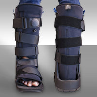
Stage I PTTD may respond to treatment that includes variations of rest. Variations in rest include an ankle brace, walking cast with an elevated heel or a hard, below-the-knee non-weight bearing cast. Pain and inflammation may be controlled with anti-inflammatory medications. It is important to be sure that Stage I patients realize that the use of shoes with additional arch support and heel elevation is imperative. Arch support and heel elevation should be continued indefinitely. Arch support, whether built into the shoe or added as an orthotic, helps support the posterior tibial tendon and decrease the amount of mechanical load applied to the posterior tibial tendon. Elevation of the heel reduces equinus, one of the most significant contributing factors to PTTD. If Stage I patients return to low heels without arch support, PTTD will recur.
Stage II patients may be treated conservatively or surgically.[9,10] Success with conservative care varies in stage II patients. Surgical correction focuses on stabilization of the subtalar joint with subtalar arthroeresis or calcaneal osteotomy. Subtalar arthroeresis is a surgical procedure that uses an implant to stabilize the subtalar joint. Subtalar arthroeresis may only be used in flexible feet. Arthroeresis is a term that means the motion of the joint is blocked without fusion. Subtalar arthroeresis can only be used in cases of Stage II posterior tibial tendonitis where mild to moderate deformation of the arch has occurred and MRI findings show the tendon to be only partially ruptured with no lengthening. Subtalar arthroeresis is typically performed in conjunction with an Achilles tendon lengthening procedure or endoscopic gastrocnemius recession to correct equinus. These procedures require casting for a period of weeks following the procedure. Alternatively, a calcaneal osteotomy or what is often referred to as a calcaneal slide procedure may also be used to stabilize the subtalar joint.
The video tab on this page shows placement of a subtalar joint implant for control of pronation in a flexible foot with PTTD. The sinus tarsi is dissected free of capsule and ligament and the implant is placed in the sinus tarsi. This procedure is completed in a hospital or outpatient surgery center using a general anesthetic. Patients can walk immediately following subtalar arthroeresis if an Achilles tendon lengthening is not performed. If an Achilles tendon lengthening is performed, a 6 week period of non-weight bearing casting is required.
When a symptomatic os tibiale externum is present, a modified Kidner procedure is performed. The following images show excision of the os tibiale externum and transposition of the posterior tibial tendon. This procedure is performed in a hospital or outpatient surgery center using a general anesthetic. Weight-bearing following the surgery is dependent upon the integrity of the tendon following excision of the os tibiale externum. Most modified Kidner procedures do require a period of non-weight bearing.


Stage III patients require stabilization of the rearfoot with procedures that structurally align the foot to be directly beneath the leg. A calcaneal osteotomy with displacement of the heel medially is commonly used in conjunction with primary repair of the posterior tibial tendon. Additionally, an Evans osteotomy with graft may be used to treat abduction of the forefoot. The Evans procedure lengthens the lateral aspect of the heel and helps to align the foot beneath the leg.
In severe cases of a rigid flatfoot that has sustained degenerative change of the subtalar joint, joint fusion (arthrodesis) is used to correct rigid deformities of the foot. These procedures are salvage procedures and require non-weight bearing casting for 8-12 weeks following surgery. A common procedure for Stage III is called a triple arthrodesis which is a technique used to fuse the subtalar joint, the talo-navicular joint and the calcaneal cuboid joint (picture at left.)
The posterior tibial tendon is reconstructed in stage III cases using a donor graft from either the long flexor tendon to the lesser toes or the long flexor tendon to the great toe. These two tendons function in the same phase of gait as the posterior tibial tendon. Therefore, grafting these tendons while maintaining their muscular origin in the lower leg works extremely well.
When to contact your doctor
PTTD is a progressive problem that should be evaluated by your podiatrist or orthopedist.
References
1. Johnson KA, Strom DE. Tibial posterior tendon dysfunction. Clin Orthop 1989;239:196-206.
2. Myerson MS. Instructional course lectures, the American Academy of Orthopedic Surgeons — Adult acquired flatfoot deformity. Treatment of dysfunction of the posterior tibial tendon. J Bone Joint Surg Am 1996;78(5):780-792.
3. Kohls-Gatzoulis J, Woods B, Angel JC, Singh D. The prevalence of symptomatic posterior tibial tendon dysfunction in women over the age of 40 in England. Foot Ankle Surg 2009;15(2):75-81.
4. Ross JA. Posterior tibial tendon dysfunction in the athlete. Clin Podiatr Med Surg 1997;14(3):479-488.
5. Rabbito M, Pohl MB, Humble N, Ferber R. biomechanical and clinical factors related to stage one posterior tibial tendon dysfunction. J Orthop Sports Phys Ther 2011;41(10):776-784.
6. Bridgeman JT, Zhang Y, Donahue H, et al. Estrogen receptor expression in posterior tibial tendon dysfunction: a pilot study. Foot Ankle Int 2010;31(12):1081-1084.
7. Alvarez RG, Marini A, Schmitt C, Saltzman CL. Stage I and II posterior tibial tendon dysfunction treated by a structured nonoperative management protocol: an orthosis and exercise program. Foot Ankle Int 2006;27(1):2-8.
8. Kulig K, Lederhaus ES, Reischl S, et al. Effect of eccentric exercise program for early tibial posterior tendinopathy. Foot Ankle Int 2009;30(9):877-885.
9. Lin JL, Balbas J, Richardson EG. Results of a non-surgical treatment of stage II posterior tibial tendon dysfunction: a 7 to 10-year follow-up. Foot Ankle Int 2008;29(8):781-786.
10. Neville CG, Lemley FR. Effect of ankle-foot orthotic devices on foot kinematics in stage II posterior tibial tendon dysfunction. Foot Ankle Int 2012;33(5):406-414.
Author(s) and date
![]() This article was written by Myfootshop.com medical advisor Jeffrey A. Oster, DPM.
This article was written by Myfootshop.com medical advisor Jeffrey A. Oster, DPM.
Competing Interests - None
Cite this article as: Oster, Jeffrey. Posterior Tibial Tendon Dysfunction. /blogs/articles/posterior-tibial-tendon-dysfunction
Most recent article update: January 14, 2021.
 Posterior Tibial Tendon Dysfunction by Myfootshop.com is licensed under a Creative Commons Attribution-NonCommercial 3.0 Unported License.
Internal reference only: ZoneM3, ZoneM6